Magnetic resonance imaging is a relatively new diagnostic method that uses the magnetic field to diagnose a myriad of diseases (many of which are undiagnosed by other imaging methods – radiographs, ultrasounds and even tomographies in some cases), using the way is made up of the human body, namely, 90% water, and how hydrogen ions respond to magnetic field stimulation.
These data are then interpreted by a computer using a special matrix, obtaining the recognizable images of the human body in the form of slices of different thicknesses in three planes (axial, coronal and sagittal with the possibility of reconstructing the image of different organs).
Sometimes it is necessary to inject intravenous contrast substance to visualize the vascular system or to characterize the vascular lesions. It is also possible to report to neighboring organs or tissues, providing preoperative information very useful.
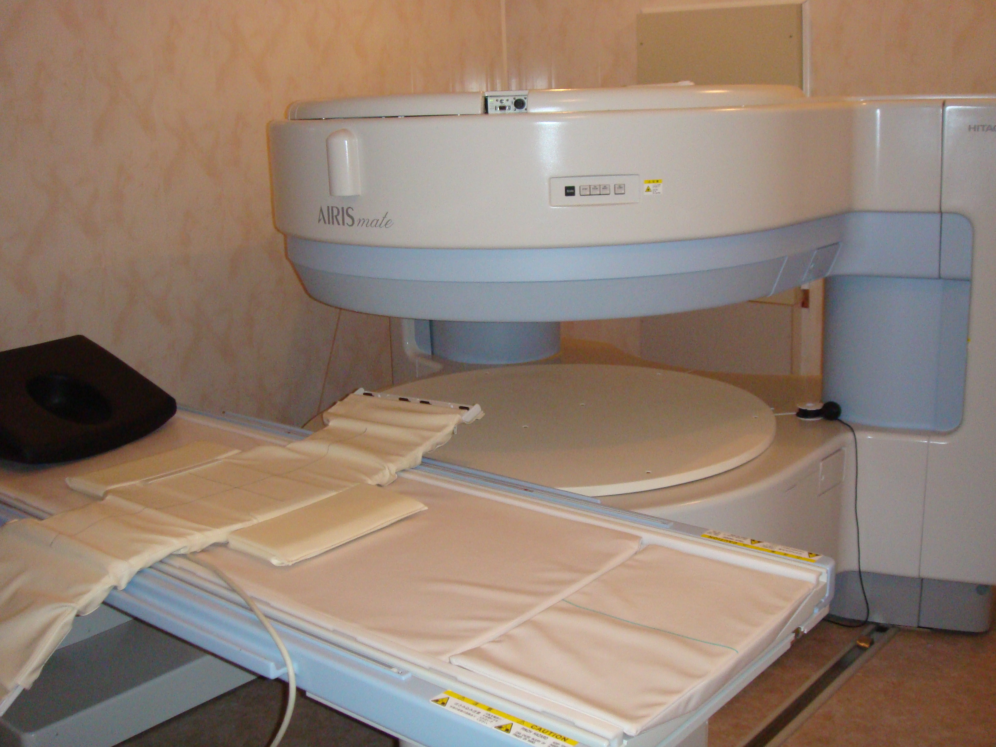
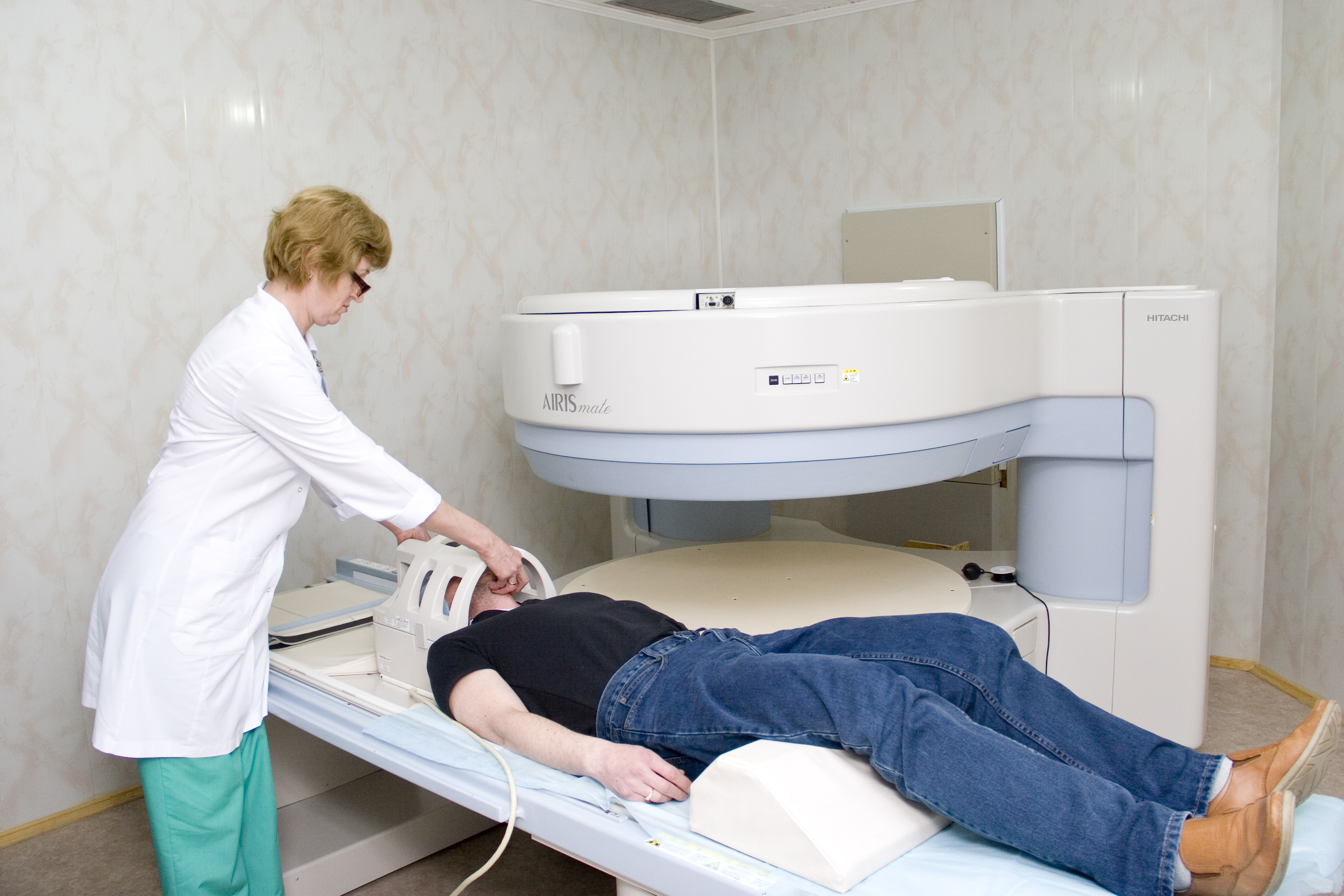
The MRI exam is done for each segment using „antennas” that concentrate the magnetic field on the exam area – for example: MRI of the spine (cervical, thoracic and lumbar), cerebral MRI, knee MRI and of course there may be combinations, adjacent regions of the human body. MRI indications include neurological and neurosurgical, orthopedic, ophthalmic, but also gynecological, gastroenterological, endocrinological and oncological pathology. It is important for the imagistic doctor to have a suspicion of diagnosis – otherwise, the examination becomes very long, and many sequences are needed to „look for” in different pathologies.